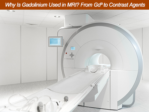
Gadolinium is used in some MRI scans because its strong paramagnetic properties change the relaxation behavior of nearby water protons. This can increase contrast on T1-weighted images and help make certain blood vessels, lesions, inflammation, and abnormal tissues easier to distinguish.
| Key question | Short answer |
| Why is gadolinium used in MRI? | It changes the relaxation behavior of nearby water protons and improves MRI contrast. |
| What does it mainly affect? | T1 relaxation and T1-weighted MRI contrast. |
| Is pure gadolinium injected? | No. Clinical MRI uses gadolinium-based contrast agents (GBCAs). |
| Does every MRI use gadolinium? | No. Many MRI scans do not require contrast. |
| Is gadolinium radioactive? | No. Its MRI function comes from its magnetic properties, not radioactivity. |
Gadolinium is a rare earth element with the chemical symbol Gd and atomic number 64. It is one of the lanthanide elements and has useful magnetic properties.
For MRI contrast, the important form is usually the Gd³⁺ ion. Gd³⁺ has seven unpaired electrons in its 4f electron shell. These unpaired electrons give it a strong magnetic moment and make gadolinium paramagnetic.
Paramagnetic materials respond to an external magnetic field. The strong paramagnetic properties of Gd³⁺ allow it to interact with nearby hydrogen nuclei.
MRI mainly detects signals from hydrogen nuclei, especially those in water and fat. By changing how these nuclei relax after radiofrequency excitation, gadolinium can change the MRI signal and improve the difference between certain tissues.
In simple terms:
Gd³⁺ → strong paramagnetism → faster proton relaxation → improved MRI contrast
The main reason gadolinium is used in MRI is its ability to change proton relaxation rates. This makes certain tissues or structures more visible on appropriately designed MRI sequences.
MRI does not directly "see" gadolinium like an ordinary X-ray image sees a metal object. Instead, MRI detects signals from hydrogen nuclei. After an MRI pulse disturbs these nuclei, they gradually return toward their normal magnetic state. This process is called relaxation.
Gadolinium's paramagnetic properties affect nearby water protons and increase their relaxation rates. The most useful effect for conventional contrast-enhanced MRI is often T1 shortening.
A shorter T1 relaxation time can produce a stronger signal on suitable T1-weighted MRI sequences. As a result, areas where the contrast agent is present may appear brighter than surrounding tissue.
This can help radiologists identify differences that might be less obvious on non-contrast images.
This is the basic scientific explanation of why gadolinium is used in MRI.
Gadolinium is not normally administered as free gadolinium ions or pure elemental metal for clinical MRI. Instead, Gd³⁺ is bound to a chelating ligand to form a gadolinium complex.
Chelation means that a molecule surrounds and binds the metal ion. In GBCAs, this chemical structure is designed to hold gadolinium within the complex during its intended use in the body.
| Material or term | What it means | Role in MRI |
| Gadolinium metal | Elemental rare earth metal, Gd | Upstream material for research and industrial applications |
| Gd³⁺ | Trivalent gadolinium ion | Paramagnetic center responsible for the MRI effect |
| Gadolinium chelate | Gd³⁺ bound to a ligand | Chemical form used to create a stable complex |
| GBCA | Gadolinium-based contrast agent | Finished pharmaceutical product used for contrast-enhanced MRI |
| Gadolinium oxide | Gd₂O₃ | Upstream rare-earth material used in research and advanced materials |
This distinction is important for both patients and material buyers. Gadolinium metal, gadolinium oxide, and a finished GBCA are different materials with different specifications and uses.
Gadolinium contrast can help improve the visualization of tissues and structures with differences in blood flow, vascularity, permeability, or other enhancement behavior.
It may be useful in several areas:
| MRI area | What contrast may help visualize |
| Brain and spine | Enhancing lesions, inflammation, and some vascular abnormalities |
| Blood vessels | Vessel anatomy and blood-flow-related abnormalities |
| Liver and abdomen | Lesion enhancement and vascular characteristics |
| Heart | Perfusion and delayed enhancement patterns |
| Musculoskeletal system | Inflammation, infection, postoperative changes, and some tumors |
| Breast and soft tissue | Enhancement patterns and vascular behavior |
The exact use depends on the clinical question, MRI protocol, patient history, and radiologist's assessment. Gadolinium does not automatically improve every MRI examination or make every abnormality easier to see.
No. Many MRI examinations are performed without gadolinium contrast.
MRI can produce detailed images using the natural differences in tissue properties. A GBCA is considered when contrast is expected to provide additional information that could help answer the clinical question.
The decision may depend on:
Therefore, the fact that gadolinium is useful in MRI does not mean that every MRI requires it.
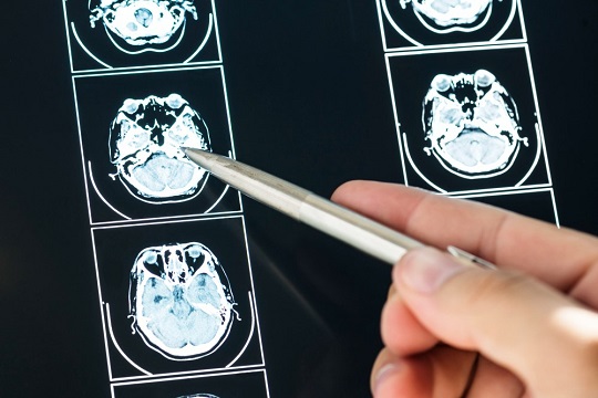
Gadolinium-based contrast agents have an established role in MRI, but they are medications, not simply imaging materials. Their use requires an assessment of the expected diagnostic benefit and patient-specific factors.
Small amounts of gadolinium can remain in the body after some GBCA administrations. The amount and duration of retention can vary among different agents.
The U.S. FDA requires warnings about gadolinium retention and states that gadolinium can remain in the body for months or years after some administrations. The FDA has also stated that, based on its review, no harmful health effects had been identified from gadolinium retention in patients with normal kidney function, while the benefits of approved GBCAs continue to outweigh potential risks.
Kidney function is an important consideration because severe renal impairment is associated with the rare but serious condition called nephrogenic systemic fibrosis (NSF).
Healthcare professionals may also consider previous contrast reactions, pregnancy, the selected GBCA, and the reason for the MRI.
Patients should tell their healthcare team about relevant medical conditions, previous contrast reactions, kidney problems, dialysis, pregnancy, and previous exposure to GBCAs.
This article provides general scientific and industry information. It does not provide medical advice. Questions about MRI contrast should be discussed with a radiologist or other qualified healthcare professional.
GBCAs are commonly classified as linear or macrocyclic, based on the structure of the ligand that binds Gd³⁺.
Linear agents use an open-chain ligand structure. Macrocyclic agents use a more constrained, ring-like structure around the gadolinium ion.
| Feature | Linear GBCAs | Macrocyclic GBCAs |
| Ligand structure | Open-chain | Ring-like or cage-like |
| Gadolinium binding | Chelated by an open-chain ligand | Held within a more constrained structure |
| Retention trend | Generally greater retention | Generally lower retention |
| Clinical selection | Depends on indication and patient factors | Depends on indication and patient factors |
The molecular structure affects the stability of the gadolinium complex and is relevant to gadolinium retention. However, macrocyclic does not mean zero risk, and patients should not choose a contrast agent themselves. The appropriate agent is selected by healthcare professionals based on the clinical situation.
For many MRI applications, a commonly used GBCA dose is around 0.1 mmol/kg, although the actual dose depends on the specific product, imaging indication, patient, and clinical protocol.
For example, at 0.1 mmol/kg, a 70 kg person would correspond to:
70 kg × 0.1 mmol/kg = 7 mmol
Using gadolinium's atomic weight of approximately 157.25 g/mol, 7 mmol corresponds to about 1,101 mg of elemental gadolinium equivalent.
This is only a simplified calculation. It is not an injection-volume recommendation. Actual GBCA concentration, volume, dose, and administration must follow the relevant product labeling and medical protocol.
The clinical product used in MRI is a finished, regulated pharmaceutical formulation. It should not be confused with elemental gadolinium metal or gadolinium oxide supplied as an upstream material.
A simplified supply chain can be viewed as:
Rare-earth source → Gadolinium separation and refining → High-purity gadolinium material → Qualified chemical intermediate → Chelated GBCA → Pharmaceutical formulation → Quality release → Clinical use
For organizations working with gadolinium upstream or in related technologies, material specifications can be critical. Requirements may include:
These requirements can vary significantly between pharmaceutical, research, ceramic, optical, magnetic, and other material applications.
AEM REE supplies rare-earth materials for industrial, scientific, research, and advanced-material applications. Its gadolinium-related offerings can include high-purity gadolinium metal, gadolinium oxide, and selected gadolinium compounds, depending on the required specification and form.
Materials may be available in forms such as powders, sheets, rods, or other customer-specified formats where applicable.
For B2B projects, important requirements may include purity, composition, physical form, quantity, packaging, and quality documentation. AEM REE works with customers on standard and customized rare-earth material requirements.
If you need high-purity gadolinium materials for research or industrial development, contact AEM REE with your required specification, form, quantity, and documentation requirements.
No. Gadolinium is used in MRI because of its paramagnetic properties, not because of radioactivity.
Gadolinium can shorten T1 relaxation time. On appropriate T1-weighted sequences, this can increase signal intensity in areas containing the contrast agent.
No. Many MRI examinations are performed without contrast. Gadolinium is used when additional contrast information is expected to be clinically useful.
No. Clinical MRI uses gadolinium-based contrast agents, in which gadolinium is chemically bound to a chelating ligand.
They differ in the structure of the ligand that binds Gd³⁺. This structural difference affects the stability of the complex and is associated with differences in gadolinium retention.
Gadolinium is used because Gd³⁺ has strong paramagnetic properties that can increase proton relaxation rates. This can improve contrast on suitable T1-weighted MRI sequences.
Gadolinium is used in MRI because its strong paramagnetic properties can change the relaxation behavior of nearby water protons, especially T1 relaxation. This effect can improve contrast and help radiologists distinguish certain tissues, blood vessels, lesions, inflammation, and other abnormalities more clearly.
However, clinical MRI does not use pure gadolinium metal. Gadolinium is incorporated into gadolinium-based contrast agents (GBCAs) through chelation, and the choice to use contrast depends on the specific imaging need and patient factors. Understanding this distinction is also important when discussing the upstream supply of gadolinium materials.
For researchers, manufacturers, and technical buyers, high-purity gadolinium metal, gadolinium oxide, and other gadolinium compounds can serve as important materials for research and advanced applications. AEM REE supports B2B customers with rare-earth materials in various purities, forms, and specifications to meet different material-development and sourcing requirements.
 +86-731-89578196